 Make sure you’ve completed the How to Use a Microscope and also the Wet Mount and Staining activities before you start here!
If you tried looking at animal cells already, you know that they wiggle and squirm all over the place. And if you tried looking when using the staining technique, you know it only makes things worse.
The heat fix technique is the one you want to use to nail your specimen to the slide and also stain it to bring out the cell structure and nuclei. This is the way scientists can look at things like bacteria.
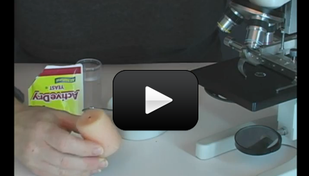
You’re going to need your microscope, slides, cover slips, eyedropper, toothpicks or tweezers, candle and matches (with adult help), stain (you can use regular iodine or Lugol’s Stain), sugar, yeast, and a container to mix your specimen in. Here’s what you do:
Please login or register to read the rest of this content.
Make sure you are logged in first or the videos will not be visible. If you still have trouble, please email me directly.
I don’t see the video on any of the experamants
No, the specimen doesn’t get washed off because you’re carefully rising it, not scrubbing it like a greasy pan. Just get most of the stain off so you can see the contrast.
Why do we rinse the slide in step 5? Does that rinse off everything we just did?
Your technique sounds good overall so I’m surprised you aren’t having better results. If you aren’t already doing so, be sure to lower the cover slip at an angle. This allows air a chance to escape which will reduce air bubbles. Also, if you are not getting much of a sample when you scrape with the toothpick, you can try with a cotton swab or Q-tip. Keep at it and good luck!
My kids and I have tried several times to view our cheek cells under the scope, only to see what I believe to be air bubbles. We are gently scraping the inside of our cheeks with a toothpick and then putting it on a slide with a drop of iodine. A small cover slide is then placed over that. I’d like to be able to find the cells and hopefully the nucleus, at the very least. Any idea what we’re doing wrong? Thank you for any help you can provide.
I’ll get a photo posted to the site so you can get a better idea… and yes, it’s very easy to kill the yeast culture if it gets above 105 degrees F, but that’s okay – you can still see great stuff as long as you didn’t destroy the cell structure.
OK. Another question. Today we did the yeast heat fix. What should we see? SHould things be moving? I am wondering if we killed our culture. We also have played with how much yeast to put on the slide as it took a while to burn off the liquid. Advice?
Never mind my last comment. Other son has a much better mouth:-).
What should a scrape from a cheek look like? We cannot seem to see anything in it? Does it need to be stained?