How does the eye work? If you are amazed as I am about how the different parts of the eye are put together, then this is the lab for you! It's important not only to learn how to take apart video cameras and blenders to find out how they work, but also to be fascinated by how the different parts of living creatures work ... like the eye!
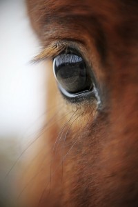
In today’s dissection, we’ll be looking at a cow eye. Because cow eyes are so similar to humans eyes, you’ll learn a lot about your own eyes by dissecting the cow eye. Eyes are a very special organ that form images from the world around you and then send the images to your brain for processing. You will be able to see the cornea, iris, pupil, connecting muscles and veins, and other features.
Materials:
[am4show have='p8;p9;p28;p55;' guest_error='Guest error message' user_error='User error message' ]
Here’s what to do:
- Take a good look at the outside of the eye. Try to find as many of the external parts as you can. You might notice the sclera, which covers the eyeball. You’ll also notice fat and muscle around the eye. Covering the front of the eye is the cornea, which was clear when the cow was alive but may look cloudy now. Next, look through the cornea to the iris (the colored part of the eye) and the iris (the dark center.)
- Cut away the fat and muscle, then use a scalpel to cut the cornea. (Get adult supervision whenever you are cutting.) The liquid that comes out is called aqueous humor. It is mostly water and helps the cornea keep its shape.
- Now make an incision into the sclera on the side opposite the cornea, and continue to cut with scissors until you end up with two halves, one with the cornea and one without.
- Place the side with the cornea on the cutting surface and cut through the cornea. You’ll hear a crunching sound. This is the sound of the many layers of tissue that make up the thick, protective cornea.
- Pull out the iris. It may be stuck to the cornea or may be back with the rest of the eye. Try to get it out in one piece. Notice that there is a hole in the center. This is the pupil, which lets in light. The pupil becomes larger or smaller to let in more or less light.
- Next, remove the lens, which looks kind of like a marble. Look through the lens and try putting it on some newspaper and looking through it to read the newspaper. What do you notice? You’ll likely see an upside down image of what you’re looking at. The lens of a cow (and human) eye, gather bits of light that bounce off an image and project those points of light as an image.
- Now go back to the rest of the eye. There may be some clear gel, called vitreous humor, in the eye. This liquid helped keep the shape of the lens. If it’s still in eye, dump it out so you can easily see the back the eye. There, you’ll see some blood vessels and a thin film. This is called the retina. When the cow looked at something, light went through the lens, and the image showed up on the retina. The retina then sent a message to the brain, through the optic nerve, and the brain interpreted what was being seen.
- If you move the retina around, you’ll find that it is only stuck to the eye in one spot. This is where the optic nerve was. If you can find the optic nerve, try pinching it with your fingers. A white substance called myelin may come out. Myelin surrounds nerves and helps messages move along more quickly.
- Behind the retina, you may find a blue-green substance called tapetum. This shiny material makes the eyes of some animals, like cows and cats, shine when light is shown on them.
Here are the basic steps to observe:
- Observe the external anatomy of the eye. See if you can locate the following:
- Sclera
- Cornea
- Optic nerve
- Excess fat and muscle tissue
- Remove the excess fat from the eye using a sharp scalpel. Then, cut through the sclera around the middle of the eye and see if you can locate the following:
- Posterior half of eye
- Optic nerve
- Retina
- Optic disc
- Choroid coat
- Tapetum lucidum
- Anterior half of eye
- Cornea
- Lens
- Iris
- Ciliary body
- Vitreous humor
- Posterior half of eye
- Cut the cornea from the eye and observe the following:
- Aqueous humor
- Cornea
- Sclera
- Iris
- Lens
- Ciliary body
[/am4show]